Activación de la microglía en el hipocampo asociada con lesión del nervio facial
Resumen
Introducción. Las lesiones del nervio facial afectan la plasticidad a largo plazo en el hipocampo, así como la memoria de reconocimiento de objetos y la memoria espacial, dos procesos dependientes de esta estructura.
Objetivo. Caracterizar en ratas el efecto de la lesión unilateral del nervio facial sobre la activación de células de la microglía en el hipocampo contralateral.
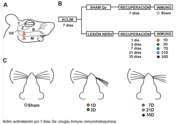
Materiales y métodos. Se hicieron experimentos de inmunohistoquímica para detectar células de la microglía en el hipocampo de ratas sometidas a lesión irreversible del nervio facial. Los animales se sacrificaron en distintos momentos después de la lesión, para evaluar la evolución de la proliferación (densidad de células) y la activación (área celular) de la microglía en el tejido del hipocampo. Los tejidos cerebrales de los animales de control se compararon con los de animales lesionados sacrificados en los días 1, 3, 7, 21 y 35 después de la lesión.
Resultados. Las células de la microglía en el hipocampo de animales con lesión del nervio facial mostraron signos de proliferación y activación a los 3, 7 y 21 días después de la lesión. Sin embargo, al cabo de cinco semanas, estas modificaciones se revirtieron, a pesar de que no hubo recuperación funcional de la parálisis facial.
Conclusiones. La lesión irreversible del nervio facial produce proliferación y activación temprana y transitoria de las células de la microglía en el hipocampo. Estos cambios podrían estar asociados con las modificaciones electrofisiológicas y las alteraciones comportamentales dependientes del hipocampo descritas recientemente.
Descargas
Referencias bibliográficas
Moran LB, Graeber MB. The facial nerve axotomy model. Brain Res Brain Res Rev. 2004;44:154-78. https://doi.org/10.1016/j.brainresrev.2003.11.004
Múnera A, Cuestas DM, Troncoso J. Peripheral facial nerve lesions induce changes in the firing properties of primary motor cortex layer 5 pyramidal cells. Neuroscience. 2012;223:140-51. https://doi.org/10.1016/j.neuroscience.2012.07.063
Urrego D, Múnera A, Troncoso J. Retracción a largo plazo del árbol dendrítico de neuronas piramidales córtico-faciales por lesiones periféricas del nervio facial. Biomédica. 2011;31:560-9. https://doi.org/10.7705/biomedica.v31i4.440
Urrego D, Troncoso J, Múnera A. Layer 5 pyramidal neurons’ dendritic remodeling and increased microglial density in primary motor cortex in a murine model of facial paralysis. Biomed Res Int. 2015;2015:482023. https://doi.org/10.1155/2015/482023
Torrado-Arévalo R, Troncoso J, Múnera A. Facial nerve axotomy induces changes on hippocampal CA3-to-CA1 long-term synaptic plasticity. Neuroscience. 2021;475:197-205. https://doi.org/10.1016/j.neuroscience.2021.08.023
Moreno C, Vivas O, Lamprea NP, Lamprea MR, Múnera A, Troncoso J. Vibrissal paralysis unveils a preference for textural rather than positional novelty in the one-trial object recognition task in rats. Behav Brain Res. 2010;211:229-35. https://doi.org/10.1016/j.bbr.2010.03.044
Patarroyo WE, García-Pérez M, Lamprea M, Múnera A, Troncoso J. Vibrissal paralysis produces increased corticosterone levels and impairment of spatial memory retrieval. Behav Brain Res. 2017;320:58-66. https://doi.org/10.1016/j.bbr.2016.11.045
Nimmerjahn A, Kirchhoff F, Helmchen F. Resting microglial cells are highly dynamic surveillants of brain parenchyma in vivo. Science. 2005;308:1314-8. https://doi.org/10.1126/science.1110647
Dávalos D, Grutzendler J, Yang G, Kim JV, Zuo Y, Jung S, et al. ATP mediates rapid microglial response to local brain injury in vivo. Nat Neurosci. 2005;8:752-8. https://doi.org/10.1038/nn1472
Ulland TK, Wang Y, Colonna M. Regulation of microglial survival and proliferation in health and diseases. Semin Immunol. 2015;27:410-5. https://doi.org/10.1016/j.smim.2016.03.011
Colonna M, Butovsky O. Microglia function in the central nervous system during health and neurodegeneration. Annu Rev Immunol. 2017;35:441-68. https://doi.org/10.1146/annurev-immunol-051116-052358
Tremblay M-È, Lowery RL, Majewska AK. Microglial interactions with synapses are modulated by visual experience. PLOS Biol. 2010;8:e1000527. https://doi.org/10.1371/journal.pbio.1000527
Wake H, Moorhouse AJ, Jinno S, Kohsaka S, Nabekura J. Resting microglia directly monitor the functional state of synapses in vivo and determine the fate of ischemic terminals. J Neurosci. 2009;29:3974-80. https://doi.org/10.1523/JNEUROSCI.4363-08.2009_15
Paolicelli RC, Bolasco G, Pagani F, Maggi L, Scianni M, Panzanelli P, et al. Synaptic pruning by microglia is necessary for normal brain development. Science. 2011;333:1456-8. https://doi.org/10.1126/science.1202529
Schafer DP, Lehrman EK, Kautzman AG, Koyama R, Mardinly AR, Yamasaki R, et al. Microglia sculpt postnatal neural circuits in an activity and complement-dependent manner. Neuron. 2012;74:691-705. https://doi.org/10.1016/j.neuron.2012.03.026
Tynan RJ, Naicker S, Hinwood M, Nalivaiko E, Buller KM, Pow DV, et al. Chronic stress alters the density and morphology of microglia in a subset of stress-responsive brain regions. Brain Behav Immun. 2010;24:1058-68. https://doi.org/10.1016/j.bbi.2010.02.001
Hinwood M, Tynan RJ, Charnley JL, Beynon SB, Day TA, Walker FR. Chronic stress induced remodeling of the prefrontal cortex: Structural re-organization of microglia and the inhibitory effect of minocycline. Cereb Cortex. 2013;23:1784-97. https://doi.org/10.1016/j.bbi.2010.02.001
Jáuregui-Huerta F, Ruvalcaba-Delgadillo Y, González-Castañeda R, García-Estrada J, González-Pérez O, Luquin S. Responses of glial cells to stress and glucocorticoids. Curr Immunol Rev. 2010;6:195-204. https://doi.org/10.2174/157339510791823790
Walker FR, Nilsson M, Jones K. Acute and chronic stress-induced disturbances of microglial plasticity, phenotype and function. Curr Drug Targets. 2013;14:1262-76. https://doi.org/10.2174/13894501113149990208
Paxinos G, Watson C. The rat brain in stereotaxic coordinates-The New Coronal Set. 6th edition. London, UK: Academic Press; 2007.
Ito D, Imai Y, Ohsawa K, Nakajima K, Fukuuchi Y, Kohsaka S. Microglia-specific localisation of a novel calcium binding protein, Iba1. Brain Res Mol Brain Res. 1998;57:1-9. https://doi.org/10.1016/s0169-328x(98)00040-0
Hill AJ. First occurrence of hippocampal spatial firing in a new environment. Exp Neurol. 1978;62:282-97. https://doi.org/10.1016/0014-4886(78)90058-4
Schmajuk NA. Role of the hippocampus in temporal and spatial navigation: An adaptive neural network. Behav Brain Res. 1990;39:205-29. https://doi.org/10.1016/0166-4328(90)90028-d
Gulli RA, Duong LR, Corrigan BW, Doucet G, Williams S, Fusi S, et al. Context-dependent representations of objects and space in the primate hippocampus during virtual navigation.Nat Neurosci. 2020;23:103-12. https://doi.org/10.1038/s41593-019-0548-3
Bird CM. The role of the hippocampus in recognition memory. Cortex. 2017;93:155-65. https://doi.org/10.1016/j.cortex.2017.05.016
Hartmann MJ, Johnson NJ, Towal RB, Assad C. Mechanical characteristics of rat vibrissae: Resonant frequencies and damping in isolated whiskers and in the awake behaving animal. J Neurosci. 2003;2:6510-9. https://doi.org/10.1523/JNEUROSCI.23-16-06510.2003
Bellistri E, Aguilar J, Brotons-Mas JR, Foffani G, Prida LM de la. Basic properties of somatosensory-evoked responses in the dorsal hippocampus of the rat. J Physiol. 2013;591:2667-86. https://doi.org/10.1113/jphysiol.2013.251892
Ren W-J, Liu Y, Zhou L-J, Li W, Zhong Y, Pang R-P, et al. Peripheral nerve injury leads to working memory deficits and dysfunction of the hippocampus by upregulation of TNF-α in rodents. Neuropsychopharmacology. 2011;36:979-92. https://doi.org/10.1038/npp.2010.236
Fasick V, Spengler RN, Samankan S, Nader ND, Ignatowski TA. The hippocampus and TNF: Common links between chronic pain and depression. Neurosci Biobehav Rev. 2015;53:139-59. https://doi.org/10.1016/j.neubiorev.2015.03.014
Fiore NT, Austin PJ. Peripheral nerve injury triggers neuroinflammation in the medial prefrontal cortex and ventral hippocampus in a subgroup of rats with coincident affective behavioural changes. Neuroscience. 2019;416:147-67. https://doi.org/10.1016/j.neuroscience.2019.08.005
Fiore NT, Austin PJ. Are the emergence of affective disturbances in neuropathic pain states contingent on supraspinal neuroinflammation? Brain Behav Immun. 2016;56:397-411. https://doi.org/10.1016/j.bbi.2016.04.012
Liu Y, Zhou L-J, Wang J, Li D, Ren W-J, Peng J, et al. TNF-α differentially regulates synaptic plasticity in the hippocampus and spinal cord by microglia-dependent mechanisms after peripheral nerve injury. J Neurosci. 2017;37:871-81. https://doi.org/10.1523/JNEUROSCI.2235-16.2016
McKenna JT, Vertes RP. Afferent projections to nucleus reuniens of the thalamus. J Comp Neurol. 2004;480:115-42. https://doi.org/10.1002/cne.20342
Vertes RP, Hoover WB, Valle ACD, Sherman A, Rodríguez JJ. Efferent projections of reuniens and rhomboid nuclei of the thalamus in the rat. J Comp Neurol. 2006;499:768-96. https://doi.org/10.1002/cne.21135
Wang Y-L, Han Q-Q, Gong W-Q, Pan D-H, Wang L-Z, Hu W, et al. Microglial activation mediates chronic mild stress-induced depressive- and anxiety-like behavior in adult rats. J Neuroinflammation. 2018;15:21. https://doi.org/10.1186/s12974-018-1054-3
Algunos artículos similares:
- Edwin Abraham Medina, Adenoma del oído medio , Biomédica: Vol. 29 Núm. 3 (2009)
- Aura Caterine Rengifo, Orlando Torres-Fernández, Disminución del número de neuronas que expresan GABA en la corteza cerebral de ratones infectados con rabia , Biomédica: Vol. 27 Núm. 4 (2007)
- Mario Francisco Guerrero, Elementos para la evaluación eficaz de productos naturales con posibles efectos antihipertensivos , Biomédica: Vol. 29 Núm. 4 (2009)
- Nina Paola Lamprea, Lina María Ortega, Gerardo Santamaría, Ladys Sarmiento, Orlando Torres-Fernández, Elaboración y evaluación de un antisuero para la detección inmunohistoquímica del virus de la rabia en tejido cerebral fijado en aldehídos , Biomédica: Vol. 30 Núm. 1 (2010)
- Diana Urrego, Alejandro Múnera, Julieta Troncoso, Retracción a largo plazo del árbol dendrítico de neuronas piramidales córtico-faciales por lesiones periféricas del nervio facial , Biomédica: Vol. 31 Núm. 4 (2011)
- Biviana Andrea Duque, Diego Aranzazu, Piedad Agudelo-Flórez, Andrés F. Londoño, Víctor H. Quiroz, Juan David Rodas, Rattus norvegicus como indicador de la circulación de Capillaria hepatica y Taenia taeniaeformis en la Plaza Minorista de Medellín, Colombia , Biomédica: Vol. 32 Núm. 4 (2012)
- Rafael A. Ulloque, Efectos de la cocaína sobre los niveles de y-aminobutirato, glutamato y aspartato en el núcleo accumbens e hipocampo de rata , Biomédica: Vol. 21 Núm. 3 (2001)
- Silvana Marisa Montenegro, María Cristina Tarrés, Juan Carlos Picena, Stella Maris Martínez, Conducta alimentaria y perfil glucémico en dos líneas de ratas con diabetes genética: eSS y eSMT. , Biomédica: Vol. 25 Núm. 4 (2005)
- Jeison Monroy-Gómez, Orlando Torres-Fernández, Distribución de calbindina y parvoalbúmina y efecto del virus de la rabia sobre su expresión en la médula espinal de ratones , Biomédica: Vol. 33 Núm. 4 (2013)
- Jorge Rivera, Marcela Neira, Edgar Parra, Jairo Méndez, Ladys Sarmiento, María Leonor Caldas, Detección de antígenos del virus del dengue en tejidos post mórtem , Biomédica: Vol. 34 Núm. 4 (2014)
| Estadísticas de artículo | |
|---|---|
| Vistas de resúmenes | |
| Vistas de PDF | |
| Descargas de PDF | |
| Vistas de HTML | |
| Otras vistas | |