Histological artifacts associated with laser and electroscalpel gingivectomy: Case series
Abstract
Introduction. Over time, efforts have been invested in the design of new instruments that overcome the disadvantages of the gold standard instrument in surgery, the scalpel. As a result, electronic equipment has emerged such as the electric scalpel and laser devices. The available evidence on these instruments suggests that the tissue response is related to each instrument’s physical and biological cutting principles.
Objective. To compare the histological changes in gingiva samples associated with surgical cutting performed with a 940 nm diode laser, a 2780 nm erbium, chromium: yttriumscandium-gallium-garnet (Er,Cr:YSGG) laser, and an electric scalpel, by presenting a series of cases.
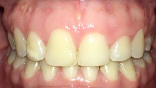
Case presentation. We present three cases of healthy patients undergoing cosmetic surgery. The clinical examination revealed exposure of a keratinized gingiva band greater than 4 mm, normal color and texture in gingival tissue, with a firm consistency and no bleeding on periodontal probing. Gingivectomy was indicated with the following protocols: Diode laser of 940 nm at 1 W, in continuous mode; Er,Cr:YSGG laser of 2780 nm at 2.5 W, 75 Hz, H mode, air 20, water 40, gold tip MT4); and electric scalpel in cutting mode at power level four. Gingival tissue samples were taken and stored in 10% formaldehyde for histological analysis.
Conclusion. All the evaluated cutting instruments generated histological changes produced by the thermal effect, the main ones being collagen coagulation and carbonization. The depth of thermal damage caused by the 2780 nm Er,Cr:YSGG laser was much lesser than that induced by the electric scalpel and the 940 nm diode laser.
Downloads
References
Storrer CL, de Oliveira ND, Deliberador TM, Ori LT, Guerrero SM, Santos FR, et al. Treatment of gingival smile: A case report. J Int Acad Periodontol. 2017;19:51-6.
Mostafa D. A successful management of sever gummy smile using gingivectomy and botulinum toxin injection: A case report. Int J Surg Case Rep. 2018;42:169-74. https://doi.org/10.1016/j.ijscr.2017.11.055
Kumar P, Rattan V, Rai S. Comparative evaluation of healing after gingivectomy with electrocautery and laser. J Oral Biol Craniofac Res. 2015;5:69-74. https://doi.org/10.1016/j.jobcr.2015.04.005
Kazakova RT, Tomov GT, Kissov CK, Vlahova AP, Zlatev SC, Bachurska SY. Histological gingival assessment after conventional and laser gingivectomy. Folia Med (Plovdiv). 2018;60:610-6. https://doi.org/10.2478/folmed-2018-0028
Kazakova R, Tomov G, Vlahova A, Zlatev S, Dimitrova M, Kazakov S, et al. Assessment of healing after diode laser gingivectomy prior to prosthetic procedures. Appl Sci (Basel). 2023;13:5527. https://doi.org/10.3390/app13095527
Mossaad AM, Abdelrahman MA, Kotb AM, Alolayan AB, Elsayed SA. Gummy smile management using diode laser gingivectomy versus botulinum toxin injection - a prospective study. Ann Maxillofac Surg. 2021;11:70-4. https://doi.org/10.4103/ams.ams_458_20
Donoso FA, Bizcar B, Sandoval C, Sandoval-Vidal P. Application of low power laser (LLLT) in pediatric patients: Review of the literature in a series of cases. Int J Odontostomat. 2018;12:269-73. https://doi.org/10.4067/S0718-381X2018000300269
Fekrazad R, Moharrami M, Chiniforush N. The esthetic crown lengthening by Er;Cr:YSGG laser: A case series. J Lasers Med Sci. 2018;9:283-7. https://doi.org/10.15171/jlms.2018.50
Nadhreen AA, Alamoudi NM, Elkhodary HM. Low-level laser therapy in dentistry: Extra-oral applications. Niger J Clin Pract. 2019;22:1313-8. https://doi.org/10.4103/njcp.njcp_53_19
Bhat P, Thakur SL, Kulkarni SS. Evaluation of soft tissue marginal stability achieved after excision with a conventional technique in comparison with laser excision: A pilot study. Indian J Dent Res. 2015;26:186-8. https://doi.org/10.4103/0970-9290.159159
Sobouti F, Rakhshan V, Chiniforush N, Khatami M. Effects of laser-assisted cosmetic smile lift gingivectomy on postoperative bleeding and pain in fixed orthodontic patients: a controlled clinical trial. Prog Orthod. 2014;15:66. https://doi.org/10.1186/s40510-014-0066-5
D’Arcangelo C, Di Nardo Di Maio F, Prosperi GD, Conte E, Baldi M, Caputi S. A preliminary study of healing of diode laser versus scalpel incisions in rat oral tissue: A comparison of clinical, histological, and immunohistochemical results. Oral Surg Oral Med Oral Pathol Oral Radiol Endod. 2007;103:764-73. https://doi.org/10.1016/j.tripleo.2006.08.002
Lione R, Pavoni C, Noviello A, Clementini M, Danesi C, Cozza P. Conventional versus laser gingivectomy in the management of gingival enlargement during orthodontic treatment: A randomized controlled trial. Eur J Orthod. 2020;42:78-85. https://doi.org/10.1093/ejo/cjz032
Cercadillo I, España A, Arnabat J, Valmaseda E, Berini L, Gay C. Histologic evaluation of thermal damage produced on soft tissues by CO2, Er,Cr:YSGG and diode lasers. Med Oral Patol Oral Cir Bucal. 2010;15:e912-8. https://doi.org/10.4317/medoral.15. e912
Abdelkarim H, Arnabat C, Parada I, Polonsky M, Arnabat J. Aerosols generation using Er,Cr:YSGG laser compared to rotary instruments in conservative dentistry: A preliminary study. J Clin Exp Dent. 2021;13:e30-6. https://doi.org/10.4317/jced.57731
Ismail A, Abushouk AI, Elmaraezy A, Menshawy A, Menshawy E, Ismail M, et al. Cutting electrocautery versus scalpel for surgical incisions: A systematic review and meta-analysis. J Surg Res. 2017;220:147-63. https://doi.org/10.1016/j.jss.2017.06.093
Jin JY, Lee SH, Yoon HJ. A comparative study of wound healing following incision with a scalpel, diode laser or Er,Cr:YSGG laser in guinea pig oral mucosa: A histological and immunohistochemical analysis. Acta Odontol Scand. 2010;68:232-8. https://doi.org/10.3109/00016357.2010.492356
Chandra GB, VinayKumar MB, Walavalkar NN, Vandana KL, Vardhan PK. Evaluation of surgical scalpel versus semiconductor diode laser techniques in the management of gingival melanin hyperpigmentation: A split-mouth randomized clinical comparative study. J Indian Soc Periodontol. 2020;24:47-53. https://doi.org/10.4103/jisp.jisp_186_19
Briceño JF, Gaviria DA, Carranza YA. Laser in Dentistry: Physical and biological foundations. Univ Odontol. 2016;35. https://doi.org/10.11144/Javeriana.uo35-75.loff 20. Arango N, Betancur N, Gómez SH. Low-level laser effect on post-exodontia inflammation of the lower third molar. Rev Nac Odontol. 2018;13:1-10. https://doi.org/10.16925/od.v13i26.1640
Verma SK, Maheshwari S, Singh RK, Chaudhari PK. Laser in dentistry: An innovative tool in modern dental practice. Natl J Maxillofac Surg. 2012;3:124-32. https://doi.org/10.4103/0975-5950.111342
Garrigo MI, Valiente C. Biological effects of low power laser radiation on tissue repair. Rev Cubana Estomatol. 1996;33:60-3.
Kaur M, Sharma YPD, Singh P, Sharma S, Wahi A. Comparative evaluation of efficacy and soft tissue wound healing using diode laser (810 nm) versus conventional scalpel technique for second-stage implant surgery. J Indian Soc Periodontol. 2018;22:228-34. https://doi.org/10.4103/jisp.jisp_46_17
Copyright (c) 2023 Biomedica

This work is licensed under a Creative Commons Attribution 4.0 International License.
| Article metrics | |
|---|---|
| Abstract views | |
| Galley vies | |
| PDF Views | |
| HTML views | |
| Other views | |


