

Description of external cuticular setae of head, thorax, legs, abdomen and genitalia of four Triatominae species
Abstract
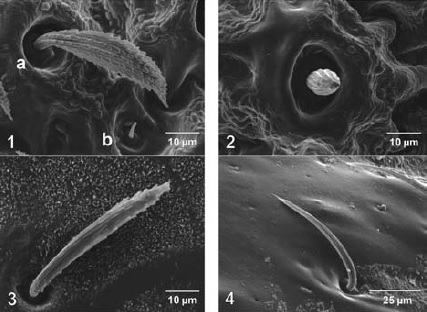
Introduction: The classification of the cuticular extensions of insects has been proposed as a taxonomic tool; however, the internal and external processes of the cuticular extensions of the Triatominae subfamily have not been fully analyzed and categorized. Objective: To describe the setae from different regions of the outer cuticle of several triatomine species by scanning electron microscopy. Materials and methods: Triatomines were washed and dried, after which different regions of the body from Eratyrus mucronatus, Triatoma maculata, Panstrongylus geniculatus and Belminus ferroae specimens were dissected, mounted on graphite double-sided adhesive tape over metal supports, metalized with gold and micrographed for further analysis. Results: We described nine types of cuticular setae. We found five types of setae in B. ferroae and T. maculata, four in P. geniculatus and only one in E. mucronatus. According to the proposed typology, type 3 seta was the most common in T. maculata, P. geniculatus and E. mucronatus, whereas type 1a predominated in B. ferroae. Conclusion: Type 3 seta was the most common in the Triatomini tribe (T. maculata, E mucronatus and P. geniculatus), whereas type 1a seta was specific to B. ferroae (tribe Bolboderini), suggesting that the surface morphology of the setae may have taxonomic value at tribe taxonomic level.
Downloads
References
Richards AG, Richards PA. The cuticular protuberances of insects. Int J Insect Morph Embryol. 1979;8:143-57.
Gorb S. Attachment devices of insect cuticle. Dordrecht: Kluwer Academic Publishers; 2002. p. 305.
Catalá S, Schofield C. Antennal sensilla of Rhodnius. J Morphol. 1994;219:193-203.
Catalá S. Antennal sensilla of Triatominae. A comparative study of five genera. Int J Insect Morphol Embriol. 1997;26: 67-73. http://dx.doi.org/10.1016/S0020-7322(97)00014-7
Rosa JA, Barata JM, Cilense M, Neto FM. Head morphology of 1st and 5th instar nymphs of Triatoma circummaculata and Triatoma rubrovaria (Hemiptera, Reduviidae). Int J Insect Morphol Embryol. 1999;28:363-75. http://dx.doi.org/10.1016/S0020-7322(99)00038-0
Rosa JA, Barata JMS, Barelli N. Morphology of abdominal bristles determined by scanning electron microscopy in six species of Triatominae (Hemiptera, Reduviidae). Mem Inst Oswaldo Cruz. 1995;90:487-8. http://dx.doi.org/10.1590/S0074-02761995000400010
Weirauch C. Hairy attachment structures in Reduviidae (Cimicomorpha, Heteroptera), with observations on the fossula spongiosa in some other Cimicomorpha. Zool Anz. 2007;246:155-75. http://dx.doi.org/10.1016/j.jcz.2007. 03.003
Catalá S. The rostral sensilla associated with eight species of Triatominae. J Morphol. 1996;228:195-201.
Altner H, Prillinger L. Ultrastructure of invertebrate chemo-, thermo-, and hygroreceptors and its functional significance. Int Rev Cytol. 1980;67:69-139. http://dx.doi.org/10.1016/S0074-7696(08)62427-4
Lent H, Wygodzinsky P. Revision of Triatominae (Hemiptera, Reduviidae) and their significance as vectors of Chagas disease. Bull Am Mus Nat Hist. 1979;163:123-520.
Catalá S, Dujardin JP. Antennal sensilla patterns indicate geographic and ecotopic variability among Triatoma infestans (Hemiptera: Reduviidae) populations. Entomol Soc Am. 200;38:423-8. http://dx.doi.org/10.1093/jmedent/38.3.423
Arroyo MA, Estaban L, Catalá S, Ángulo VM. Variación del fenotipo antenal de poblaciones del domicilio, peridomicilio y silvestres de Triatoma dimidiata (Hemiptera: Reduviidae) en Santander, Colombia. Biomédica. 2007;27(Supl.1):92-100. http://dx.doi.org/10.7705/biomedica.v27i1.252
Weirauch C. Pretarsal structures in Reduviidae (Heteroptera, Insecta). Acta Zool Stockh. 2005;86:91-110.
Some similar items:
- Kevin Escandón-Vargas, Carlos A. Muñoz-Zuluaga, Liliana Salazar, Blood-feeding of Rhodnius prolixus , Biomedica: Vol. 37 No. 3 (2017)
- Marlene Reyes, Víctor Manuel Angulo, Life cycle of Triatoma dimidiata Latreille, 1811 (Hemiptera, Reduviidae) under laboratory conditions: production of nymphs for biological tests , Biomedica: Vol. 29 No. 1 (2009)
- Piedad Agudelo-Flórez, Marcos Restrepo, Natalí Moreno, Diagnosis of leptospirosis by dark-field microscopy of blood samples and culture , Biomedica: Vol. 28 No. 1 (2008)
- José Alejandro Martínez-Ibarra, Jorge Alejandro Martínez-Grant, Miguel Roberto Verdugo-Cervantes, Rafael Bustos-Saldaña, Benjamín Nogueda-Torres, Monitoring triatomid bug (Hemiptera: Reduviidae) presence by sentinel chicken coops in Southern Jalisco State, México , Biomedica: Vol. 30 No. 1 (2010)
- María Imaz, Sonia Allassia, Mónica Aranibar, Alba Gunia, Susana Poggi, Ana Togneri, Lidia Wolff, Group of Implementation of Fluorescence, Performance of LED fluorescence microscopy for the detection of acid-fast bacilli from respiratory samples in peripheral laboratories in Argentina , Biomedica: Vol. 37 No. 2 (2017)
- Nelson Grisales, Omar Triana, Víctor Angulo, Nicolás Jaramillo, Gabriel Parra-Henao, Francisco Panzera, Andrés Gómez-Palacio, Genetic differentiation of three Colombian populations of Triatoma dimidiata (Heteroptera: Reduviidae) by ND4 mitochondrial gene molecular analysis , Biomedica: Vol. 30 No. 2 (2010)
- Marlene Reyes, Víctor Manuel Angulo, Claudia Magaly Sandoval, Toxic effect of b-cipermethrin, deltamethrin and fenitrothion in colonies of Triatoma dimidiata (Latreille, 1811) and Triatoma maculata (Erichson, 1848) (Hemiptera, Reduviidae) , Biomedica: Vol. 27 No. 1esp (2007): Enfermedad de Chagas
- Corina María Arroyo, Lyda Esteban, Silvia Catalá, Víctor Manuel Angulo, Antennal phenotype variation in sylvatic, peridomestic and domestic populations of Triatoma dimidiata (Hemiptera: Reduviidae) from Santander, Colombia. , Biomedica: Vol. 27 No. 1esp (2007): Enfermedad de Chagas
- Andrea Arévalo, Julio César Carranza, Felipe Guhl, Gustavo Adolfo Vallejo, Electrophoretic patterns of salivary hemeproteins (nitrophorines) of Rhodnius colombiensis and R. prolixus (Hemiptera, Reduviidae, Triatominae) , Biomedica: Vol. 27 No. 1esp (2007): Enfermedad de Chagas
- Felipe Guhl, Germán Aguilera, Néstor Pinto, Daniela Vergara, Updated geographical distribution and ecoepidemiology of the triatomine fauna (Reduviidae: Triatominae) in Colombia , Biomedica: Vol. 27 No. 1esp (2007): Enfermedad de Chagas
| Article metrics | |
|---|---|
| Abstract views | |
| Galley vies | |
| PDF Views | |
| HTML views | |
| Other views | |


