

Inmunohistochemical detection of pandemic SARSCoV- 2 antigens in lung tissue
Abstract
The COVID-19 pandemic caused by the SARS-CoV-2 virus has generated globally more than 110.7 million infections and 2.4 million deaths. The severity of this infection can range from asymptomatic, mild to severe.
To know the possible associations between the presence of the virus and histopathological alterations found in tissues of fatal cases of COVID-19, the presence of the virus in the lung tissue of a patient with a clinical history of SARS-CoV-2 infection was evaluated.
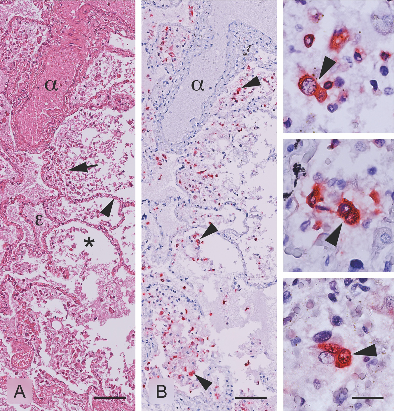
Lung tissue was histologically processed for immunohistochemical detection of SARSCoV-2. In the histopathological study, morphological changes associated with pneumonitis of viral origin were observed. Likewise, the location of the SARS-CoV-2 virus was observed mainly in the cytoplasm of the cells of the inflammatory infiltrate.
Downloads
References
Park SE. Epidemiology, virology, and clinical features of severe acute respiratory syndromecoronavirus-2 (SARS-CoV-2; Coronavirus Disease-19). Clin Exp Pediatr. 2020;63:119-24. https://doi.org/10.3345/cep.2020.00493
World Health Organization. COVID-19 Weekly Epidemiological Update 2021. Accessed: February 23, 2021. Available at: https://www.who.int/publications/m/item/weeklyepidemiological-update---23-february-2021
Álvarez-Díaz DA, Franco-Muñoz C, Laiton-Donato K, Usme-Ciro JA, Franco-Sierra ND, Flórez-Sánchez AC, et al. Molecular analysis of several in-house rRT-PCR protocols for SARS-CoV-2 detection in the context of genetic variability of the virus in Colombia. Infect Genet Evol. 2020;84. https://doi.org/10.1016/j.meegid.2020.104390
Instituto Nacional de Salud. COVID-19 en Colombia. Accessed: February 25, 2021. Available at: https://www.ins.gov.co/Noticias/Paginas/coronavirus-casos.aspx
Belouzard S, Millet JK, Licitra BN, Whittaker GR. Mechanisms of coronavirus cell entry mediated by the viral spike protein. Viruses. 2012;4:1011-33. https://doi.org/10.3390/v4061011
Zhang H, Penninger JM, Li Y, Zhong N, Slutsky AS. Angiotensin-converting enzyme 2 (ACE2) as a SARS-CoV-2 receptor: Molecular mechanisms and potential therapeutic target. Intensive Care Med. 2020;46:586-90. https://doi.org/10.1007/s00134-020-05985-9
Ding Y, He L, Zhang Q, Huang Z, Che X, Hou J, et al. Organ distribution of severe acute respiratory syndrome (SARS) associated coronavirus (SARS-CoV) in SARS patients: Implications for pathogenesis and virus transmission pathways. J Pathol. 2004;203:622-30. https://doi.org/10.1002/path.1560
Farcas GA, Poutanen SM, Mazzulli T, Willey BM, Butany J, Asa SL, et al. Fatal severe acute respiratory syndrome is associated with multiorgan involvement by coronavirus. J Infect Dis. 2005;191:193-7. https://doi.org/10.1086/426870
Satija N, Lal SK. The molecular biology of SARS coronavirus. Ann N Y Acad Sci. 2007;1102:26-38. https://doi.org/10.1196/annals.1408.002
Martines R, Ritter J, Matkovic E, Gary J, Bollweg B, Bullock H, et al. Pathology and pathogenesis of SARS-CoV-2 associated with fatal coronavirus disease, United States. Emerg Infect Dis. 2020;26:2005. https://doi.org/10.3201/eid2609.202095
Chu H, Chan JF-W, Yuen TT-T, Shuai H, Yuan S, Wang Y, et al. Comparative tropism, replication kinetics, and cell damage profiling of SARS-CoV-2 and SARS-CoV with implications for clinical manifestations, transmissibility, and laboratory studies of COVID-19: An observational study. Lancet Microbe. 2020;1:e14-e23. https://doi.org/10.1016/s2666-5247(20)30004-5
Wang D, Hu B, Hu C, Zhu F, Liu X, Zhang J, et al. Clinical characteristics of 138 hospitalized patients with 2019 novel coronavirus-infected pneumonia in Wuhan, China. JAMA. 2020;323:1061-9. https://doi.org/10.1001/jama.2020.1585
Vaira LA, Salzano G, Deiana G, De Riu G. Anosmia and ageusia: Common findings in COVID-19 patients. Laryngoscope. 2020;130:1787. https://doi.org/10.1002/lary.28692
Menter T, Haslbauer JD, Nienhold R, Savic S, Hopfer H, Deigendesch N, et al. Postmortem examination of COVID-19 patients reveals diffuse alveolar damage with severe capillary congestion and variegated findings in lungs and other organs suggesting vascular dysfunction. Histopathology. 2020;77:198-209. https://doi.org/10.1111/his.14134
Tian S, Xiong Y, Liu H, Niu L, Guo J, Liao M, et al. Pathological study of the 2019 novel coronavirus disease (COVID-19) through postmortem core biopsies. Mod Pathol. 2020;33:1007-14. https://doi.org/10.1038/s41379-020-0536-x
Griffin DO, Jensen A, Khan M, Chin J, Chin K, Saad J, et al. Pulmonary embolism and increased levels of d-Dimer in patients with coronavirus disease. Emerg Infect Dis. 2020;26:1941-3. https://doi.org/10.3201/eid2608.201477
Mohanty SK, Satapathy A, Naidu MM, Mukhopadhyay S, Sharma S, Barton LM, et al. Severe acute respiratory syndrome coronavirus-2 (SARS-CoV-2) and coronavirus disease 19 (COVID-19) - anatomic pathology perspective on current knowledge. Diagn Pathol. 2020;15:103. https://doi.org/10.1186/s13000-020-01017-8
Some similar items:
- Edwin Abraham Medina, Middle ear adenoma , Biomedica: Vol. 29 No. 3 (2009)
- Aura Caterine Rengifo, Orlando Torres-Fernández, Decreased number neurons expressing GABA in the cerebral cortex of rabies-infected mice , Biomedica: Vol. 27 No. 4 (2007)
- Juan Carlos Miguel, Ariana Erazo, Fernanda Beduino, Juan Carlos Picena, María Isabel Luciano, Gustavo Pizzuti, María Cristina Tarrés, Silvana Montenegro, Stella Maris Martínez, Chronic bronchial dilatations in different colonies of laboratory rats , Biomedica: Vol. 22 No. 2 (2002)
- Henry A. Vargas, Martín Rondón, Rodolfo Dennis, Pharmacological treatment and impairment of pulmonary function in patients with type 2 diabetes: a cross-sectional study , Biomedica: Vol. 36 No. 2 (2016)
- Ana Madeleine Barrera, Leslie Vargas, Idiopathic pulmonary hemosiderosis with dendriform pulmonary ossification , Biomedica: Vol. 36 No. 4 (2016)
- Gerardo Santamaria, Jeison Monroy-Gómez, Orlando Torres-Fernández, Neuroanatomical evidence of the transport of the rabies virus through the propriospinal tract in the spinal cord of mice , Biomedica: Vol. 38 No. 2 (2018)
- Julián Alfredo Fernández-Niño , Andrés Cubillos-Novella, Ietza Bojórquez , Michael Rodríguez , Recommendations for the response against COVID-19 in migratory contexts under a closed border: The case of Colombia , Biomedica: Vol. 40 No. Supl. 2 (2020): SARS-CoV-2 y COVID-19
- Jeimmy Cerón, Julieta Troncoso, Facial nerve injury-associated hippocampal microglial activation , Biomedica: Vol. 42 No. 1 (2022)
- Claudia Milena Cuéllar-Segura, Lessons learned in public health from COVID-19: A look towards future epidemics and pandemics , Biomedica: Vol. 42 No. Sp. 2 (2022): Covid-19
- Jorge Rivera , Sheryll Corchuelo, Julián Naizaque, Édgar Parra, Eugenio Aladino Meek, Diego Álvarez-Díaz, Marcela Mercado , Orlando Torres-Fernández, Histopathology of COVID-19: An illustration of the findings from fatal cases , Biomedica: Vol. 43 No. 1 (2023)
Copyright (c) 2022 Biomedica

This work is licensed under a Creative Commons Attribution 4.0 International License.
| Article metrics | |
|---|---|
| Abstract views | |
| Galley vies | |
| PDF Views | |
| HTML views | |
| Other views | |


