Estrés oxidativo en células endoteliales inducido por el suero de mujeres con diferentes manifestaciones clínicas del síndrome antifosfolípido
Resumen
Introducción. El síndrome antifosfolípido se caracteriza por la presencia persistente de anticuerpos antifosfolípidos y manifestaciones clínicas de trombosis o morbilidad gestacional, las cuales se asocian con estrés oxidativo y disfunción endotelial.
Objetivo. Evaluar los marcadores de estrés oxidativo en células endoteliales, inducidos por el suero de mujeres con diferentes manifestaciones clínicas del síndrome antifosfolípido y analizar la capacidad antioxidante de los sueros.
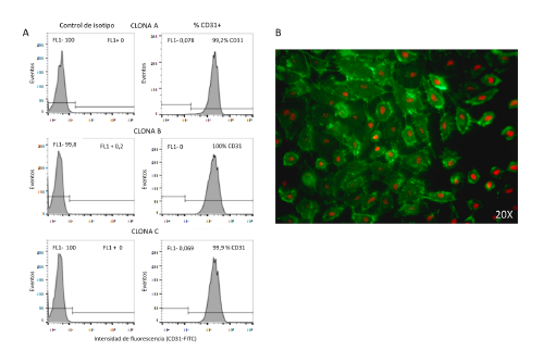
Materiales y métodos. Se incluyeron 48 mujeres que fueron clasificadas así: presencia de anticuerpos antifosfolípidos y criterios clínicos de morbilidad gestacional, trombosis vascular o ambas. Como grupos control se incluyeron mujeres negativas para anticuerpos antifosfolípidos. En un modelo in vitro de células endoteliales estimuladas con los sueros de las mujeres del estudio, se determinaron algunos marcadores de estrés oxidativo por citometría de flujo. También, se analizó la capacidad antioxidante de los sueros incluidos.
Resultados. Los sueros de los grupos de mujeres con síndrome antifosfolípido que presentaban trombosis, con morbilidad gestacional o sin ella, generaron un incremento significativo (p<0,05 y p<0,001) en los marcadores de estrés oxidativo endotelial, en contraste con el control de suero humano normal. No se observaron diferencias en el efecto de los sueros de los diferentes grupos de estudio sobre la lipoperoxidación endotelial. Tampoco se encontró diferencia en la actividad antioxidante de los sueros.
Conclusión. El estrés oxidativo mitocondrial en el endotelio se asocia con la presencia de trombosis. Sin embargo, cuando esta se asocia con morbilidad gestacional, también se genera estrés oxidativo intracelular
Descargas
Referencias bibliográficas
Miyakis S, Lockshin MD, Atsumi T, Branch DW, Brey RL, Cervera R, et al. International consensus statement on an update of the classification criteria for definite antiphospholipid syndrome (APS). J Thromb Haemost. 2006;4:295-306. https://doi.org/10.1111/j.1538-7836.2006.01753.x
Esteve-Valverde E, Ferrer-Oliveras R, Alijotas-Reig J. Obstetric antiphospholipid syndrome. Rev Clin Esp. 2016;216:135-45. https://doi.org/10.1016/j.rce.2015.09.003
Cervera R, Serrano R, Pons-Estel GJ, Ceberio-Hualde L, Shoenfeld Y, de Ramon E, et al. Morbidity and mortality in the antiphospholipid syndrome during a 10-year period: A multicentre prospective study of 1000 patients. Ann Rheum Dis. 2015;74:1011-8. https://doi.org/10.1136/annrheumdis-2013-204838
Meroni PL, Borghi MO, Raschi E, Tedesco F. Pathogenesis of antiphospholipid syndrome: Understanding the antibodies. Nat Rev Rheumatol. 2011;7:330-9. https://doi.org/10.1038/nrrheum.2011.52
Chighizola CB, Raschi E, Borghi MO, Meroni PL. Update on the pathogenesis and treatment of the antiphospholipid syndrome. Curr Opin Rheumatol. 2015;27:476-82.
https://doi.org/10.1097/BOR.0000000000000200
Ripoll VM, Pregnolato F, Mazza S, Bodio C, Grossi C, McDonnell T, et al. Gene expression profiling identifies distinct molecular signatures in thrombotic and obstetric antiphospholipid syndrome. J Autoimmun. 2018;93:114-23. https://doi.org/10.1016/j.jaut.2018.07.002
Álvarez AM, Balcázar N, San Martín S, Markert UR, Cadavid AP. Modulation of antiphospholipid antibodies-induced trophoblast damage by different drugs used to prevent pregnancy morbidity associated with antiphospholipid syndrome. Am J Reprod Immunol. 2017;77:77(4):e12634. https://doi.org/10.1111/aji.12634
Velásquez M, Álvarez ÁM, Cadavid ÁP. Cuantificación sistematizada de la remodelación vascular in vitro en la morbilidad gestacional asociada al síndrome antifosfolípido. Rev Chil Obstet Ginecol. 2016;81:455-64 https://doi.org/10.4067/S0717-75262016000600002
Sacharidou A, Shaul PW, Mineo C. New insights in the pathophysiology of antiphospholipid syndrome. Semin Thromb Hemost. 2018;44:475-82. https://doi.org/10.1055/s-0036-1597286
López-Pedrera C, Barbarroja N, Jiménez-Gómez Y, Collantes-Estévez E, Aguirre MA, Cuadrado MJ. Oxidative stress in the pathogenesis of atherothrombosis associated with anti-phospholipid syndrome and systemic lupus erythematosus: New therapeutic approaches. Rheumatology (Oxford). 2016;55:2096-108.
https://doi.org/10.1093/rheumatology/kew054
Lum H, Roebuck KA. Oxidant stress and endothelial cell dysfunction. Am J Physiol Cell Physiol. 2001;280:C719-41. https://doi.org/10.1152/ajpcell.2001.280.4.C719
Simoncini S, Sapet C, Camoin-Jau L, Bardin N, Harle JR, Sampol J, et al. Role of reactive oxygen species and p38 MAPK in the induction of the pro-adhesive endothelial state mediated by IgG from patients with anti-phospholipid syndrome. Int Immunol. 2005;17:489-500. https://doi.org/10.1093/intimm/dxh229
Pierangeli SS, Espinola RG, Liu X, Harris EN. Thrombogenic effects of antiphospholipid antibodies are mediated by intercellular cell adhesion molecule-1, vascular cell adhesion molecule-1, and P-selectin. Circ Res. 2001;88:245-50
Kojda G, Harrison D. Interactions between NO and reactive oxygen species: Pathophysiological importance in atherosclerosis, hypertension, diabetes and heart failure. Cardiovasc Res. 1999;43:562-71.
Lubos E, Handy DE, Loscalzo J. Role of oxidative stress and nitric oxide in atherothrombosis. Front Biosci. 2008;13:5323-44.
Giannakopoulos B, Krilis SA. The pathogenesis of the antiphospholipid syndrome. N Engl J Med. 2013;368:1033-44. https://doi.org/10.1056/NEJMra1112830
Ioannou Y, Zhang JY, Qi M, Gao L, Qi JC, Yu DM, et al. Novel assays of thrombogenic pathogenicity in the antiphospholipid syndrome based on the detection of molecular oxidative modification of the major autoantigen beta2-glycoprotein I. Arthritis Rheum. 2011;63:2774-82. https://doi.org/10.1002/art.30383
Pérez-Sánchez C, Barbarroja N, Messineo S, Ruiz-Limón P, Rodríguez-Ariza A, Jiménez-Gómez Y, et al. Gene profiling reveals specific molecular pathways in the pathogenesis of atherosclerosis and cardiovascular disease in antiphospholipid syndrome, systemic lupus erythematosus and antiphospholipid syndrome with lupus. Ann Rheum Dis. 2015;74:1441-9. https://doi.org/10.1136/annrheumdis-2013-204600
Kwak JY, Gilman-Sachs A, Beaman KD, Beer AE. Autoantibodies in women with primary recurrent spontaneous abortion of unknown etiology. J Reprod Immunol. 1992;22:15-31. https://doi.org/10.1016/0165-0378(92)90003-M
Robinson KM, Janes MS, Beckman JS. The selective detection of mitochondrial superoxide by live cell imaging. Nat Protoc. 2008;3:941. https://doi.org/10.1038/nprot.2008.56
Drummen GP, Makkinje M, Verkleij AJ, den Kamp JAO, Post JA. Attenuation of lipid peroxidation by antioxidants in rat-1 fibroblasts: Comparison of the lipid peroxidation reporter molecules cis-parinaric acid and C11-BODIPY 581/591 in a biological setting. Biochim Biophys Acta. 2004;1636:136-50. https://doi.org/10.1016/j.bbalip.2003.10.013
Nicolescu AC, Li Q, Brown L, Thatcher GR. Nitroxidation, nitration, and oxidation of a BODIPY fluorophore by RNOS and ROS. Nitric Oxide. 2006;15:163-76. https://doi.org/10.1016/j.niox.2006.01.010
Mackness MI, Mackness B, Durrington PN, Connelly PW, Hegele RA. Paraoxonase: Biochemistry, genetics and relationship to plasma lipoproteins. Curr Opin Lipidol 1996;7:69-76.
Furlong CE, Richter RJ, Seidel SL, Costa LG, Motulsky AG. Spectrophotometric assays for the enzymatic hydrolysis of the active metabolites of chlorpyrifos and parathion by plasma paraoxonase/arylesterase. Anal Biochem. 1989;180:242-7. https://doi.org/10.1016/0003-2697(89)90424-7
MacDonald-Wicks LK, Wood LG, Garg ML. Methodology for the determination of biological antioxidant capacity in vitro: A review. J Sci Food Agric. 2006;86:2046-56. https://doi.org/10.1002/jsfa.2603
Serafini M, Maiani G, Ferro-Luzzi A. Alcohol-free red wine enhances plasma antioxidant capacity in humans. J Nutr. 1998;128:1003-7. https://doi.org/10.1093/jn/128.6.1003
Bertolaccini ML, Amengual O, Atsumi T, Binder WL, de Laat B, Forastiero R, et al. 'Non-criteria' aPL tests: Report of a task force and preconference workshop at the 13th International Congress on Antiphospholipid Antibodies, Galveston, TX, USA, April 2010. Lupus. 2011;20:191-205. https://doi.org/10.1177/0961203310397082
Álvarez AM, Mulla MJ, Chamley LW, Cadavid AP, Abrahams VM. Aspirin-triggered lipoxin prevents antiphospholipid antibody effects on human trophoblast migration and endothelial cell interactions. Arthritis Rheumatol. 2015;67:488-97. https://doi.org/10.1002/art.38934
Redecha P, Franzke CW, Ruf W, Mackman N, Girardi G. Neutrophil activation by the tissue factor/Factor VIIa/PAR2 axis mediates fetal death in a mouse model of antiphospholipid syndrome. J Clin Invest. 2008;118:3453-61. https://doi.org/10.1172/JCI36089
Weiler H. Tracing the molecular pathogenesis of antiphospholipid syndrome. J Clin Invest. 2008;118:3276-8. https://doi.org/10.1172/JCI37243
Wu F, Tian FJ, Lin Y. Oxidative stress in placenta: Health and diseases. Biomed Res Int. 2015;2015:293271. https://doi.org/10.1155/2015/293271
Meroni PL, Borghi MO, Grossi C, Chighizola CB, Durigutto P, Tedesco F. Obstetric and vascular antiphospholipid syndrome: Same antibodies but different diseases? Nat Rev Rheumatol. 2018;14:433-40. https://doi.org/10.1038/s41584-018-0032-6
Prinz N, Clemens N, Canisius A, Lackner KJ. Endosomal NADPH-oxidase is critical for induction of the tissue factor gene in monocytes and endothelial cells. Lessons from the antiphospholipid syndrome. Thromb Haemost. 2013;109:525-31. https://doi.org/10.1160/TH12-06-0421
Dikalov SI, Nazarewicz RR, Bikineyeva A, Hilenski L, Lassegue B, Griendling KK, et al. Nox2-induced production of mitochondrial superoxide in angiotensin II-mediated endothelial oxidative stress and hypertension. Antioxid Redox Signal. 2014;20:281-94. https://doi.org/10.1089/ars.2012.4918
Roubey RA. Tissue factor pathway and the antiphospholipid syndrome. J Autoimmun. 2000;15:217-20. https://doi.org/10.1006/jaut.2000.0397
Benhamou Y, Miranda S, Armengol G, Harouki N, Drouot L, Zahr N, et al. Infliximab improves endothelial dysfunction in a mouse model of antiphospholipid syndrome: Role of reduced oxidative stress. Vascul Pharmacol. 2015;71:93-101. https://doi.org/10.1016/j.vph.2015.03.014
Sacharidou A, Chambliss KL, Ulrich V, Salmon JE, Shen YM, Herz J, et al. Antiphospholipid antibodies induce thrombosis by PP2A activation via apoER2-Dab2-SHC1 complex formation in endothelium. Blood. 2018;131:2097-110. https://doi.org/10.1182/blood-2017-11-814681
Delgado-Alves J, Ames P, Donohue S, Stanyer L, Noorouz-Zadeh J, Ravirajan C, et al. Antibodies to high-density lipoprotein and β2-glycoprotein I are inversely correlated with paraoxonase activity in systemic lupus erythematosus and primary antiphospholipid syndrome. Arthritis Rheum. 2002;46:2686-94. https://doi.org/10.1002/art.10542
Delgado- Alves J, Mason L, Ames P, Chen P, Rauch J, Levine J, et al. Antiphospholipid antibodies are associated with enhanced oxidative stress, decreased plasma nitric oxide and paraoxonase activity in an experimental mouse model. Rheumatology (Oxford). 2005;44:1238-44. https://doi.org/10.1093/rheumatology/keh722
Fenzl V, Flegar-Mestric Z, Perkov S, Andrisic L, Tatzber F, Zarkovic N, et al. Trace elements and oxidative stress in hypertensive disorders of pregnancy. Arch Gynecol Obstet. 2013;287:19-24. https://doi.org/10.1007/s00404-012-2502-4
Algunos artículos similares:
- Edisson Rodríguez, Aura María Gil-Villa, Daniel Camilo Aguirre-Acevedo, Walter Cardona-Maya, Ángela P. Cadavid, Evaluación de parámetros seminales no convencionales en individuos cuyas parejas presentan muerte embrionaria temprana recurrente: en busca de un valor de referencia , Biomédica: Vol. 31 Núm. 1 (2011)
- Luis Miguel Sosa, Luz Libia Cala, Julio César Mantilla, Tuberculosis congénita asociada con tuberculosis materna miliar diseminada , Biomédica: Vol. 27 Núm. 4 (2007)
- Claudia Ayala, Reggie García, Edith Cruz, Karol Prieto, Marta Bermúdez, Niveles de homocisteína y polimorfismos de los genes de la MTHFR y la CBS en pacientes colombianos con trombosis venosa superficial y profunda , Biomédica: Vol. 30 Núm. 2 (2010)
- Germán González, Marta Lía Valencia, Nelson Armando Agudelo, Liliana Acevedo, Isabel Cristina Vallejo, Morbilidad sentida de las urgencias médicas y la utilización de los servicios de salud en Medellín, Colombia, 2005-2006 , Biomédica: Vol. 27 Núm. 2 (2007)
- Jairo Lizarazo, Melva Linares, Catalina de Bedout, Ángela Restrepo, Clara Inés Agudelo, Elizabeth Castañeda, Grupo Colombiano para el Estudio de la Criptococosis, Estudio clínico y epidemiológico de la criptococosis en Colombia: resultados de nueve años de la encuesta nacional, 1997-2005 , Biomédica: Vol. 27 Núm. 1 (2007)
- Berta Nelly Restrepo, María Teresa Restrepo, Juan Camilo Beltrán, Mónica Rodríguez, Ruth Emilia Ramírez, Estado nutricional de niños y niñas indígenas de hasta seis años de edad en el resguardo Embera-Katío, Tierralta, Córdoba, Colombia , Biomédica: Vol. 26 Núm. 4 (2006)
- Jorge Andrés Jiménez, Lady Diana Ladino, Carlos Santiago Uribe, Alejandro Guerra, Juan Diego Ciro, Olga Elena Hernández, Jorge Andrés Ochoa, Neurosífilis meningovascular con trombosis de la arteria basilar , Biomédica: Vol. 32 Núm. 1 (2012)
- Helena Hernández-Cuervo, Solangy Usme, Juan José Yunis, Genotipos frecuentemente asociados a trombofilias , Biomédica: Vol. 34 Núm. 1 (2014)
- Stella Carrasco Rodríguez, Myriam Sanchez Gómez, Niveles séricos de IGF-I e IGFBP-3 en adolescentes embarazadas provenientes de un sector socioeconómico de bajos ingresos , Biomédica: Vol. 21 Núm. 4 (2001)
- Diana Carolina Cáceres, Vilma Fabiola Izquierdo, Leonardo Mantilla, Jorge Jara, Martha Velandia, Perfil epidemiológico de la población desplazada por el conflicto armado interno del país en un barrio de Cartagena, Colombia, 2000. , Biomédica: Vol. 22 (2002): Suplemento 2
| Estadísticas de artículo | |
|---|---|
| Vistas de resúmenes | |
| Vistas de PDF | |
| Descargas de PDF | |
| Vistas de HTML | |
| Otras vistas | |